
We offer a wide range of services to meet the needs of our horse owners. We offer both in clinic as well as on the farm services.
- Lameness Diagnostics
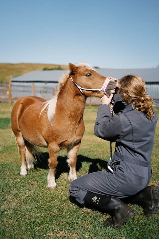
- Dentistry
- Ultrasound
- Digital radiographs
- Coggins and Health Inspections
- Vaccination and Deworming
- Basic Surgery
- Castration
- Medical Colic Treatment
- Foal Care
- Pre-purchase/Insurance Exams
- Wound Care
- Sick Animal Care and Diagnostics (Urinalysis, Parasitology, Radiology, and Blood Chemistry)
- Euthanasia
- Laser Therapy
Equine Preventable Diseases
Eastern & Western Encephalomyelitis
Viral; birds are the reservoir hose and mosquitoes are the vector (responsible for transmitting the disease). Horses and humans are dead-end hosts - mosquitoes cannot spread the virus from them to other horses or humans. Clinical signs: lethargy, depression, fever, ataxia (lack of voluntary coordination of muscle movements), anorexia, paralysis, circling, head pressing, hyper-excitability, blindness, and seizures. If the animal survives they may have permanent CNS damage (behavioural and learning deficits). Treatment: none; can provide supportive care. Prevention: vaccination at the beginning of mosquito season each year (end of May/beginning of June).
West Nile Virus Disease
Viral; transmitted between vertebrates by certain mosquitoes. The virus attacks the brain and spinal cord. Birds are considered the main vertebrate host, and other mammals are considered dead-end hosts. The virus can infect any mammal, but the main clinical signs are seen in birds, horses, and humans. Clinical signs: there may be no signs at all, mild signs, or fatal; approximately 1/3 of infected horses die from the disease. Low-grade fever, lack of appetite, depression, colic, ataxia, recumbency, seizures, and personality changes that range from hyper-excitability to apprehensiveness, unresponsiveness, and coma. Some horses develop paralysis and have to be euthanized, others die spontaneously. Many horses start to show signs of recovery within 3 to 7 days and complete a full recovery within 1 to 6 months. Treatment: none; can provide supportive care. Prevention: vaccination at the beginning of mosquito season each year (end of May/beginning of June).
Rabies
Viral; causes lethal polioencephalomyelitis and ganglionitis in all warm-blooded mammals. It is a reportable disease in Canada. Bats are the only carriers that can show symptoms or be symptomless carriers. The virus is spread from the carrier animal though the saliva. Once the virus gains entrance into the body, it multiplies at the local site, and then enters the nervous system at the motor end plates of the local nerves. From there it moves to the spinal cord and then the brain. Clinical signs: two forms: furious or paralytic. Clinical signs in the furious form include tense, alert appearance, hypersensitivity to sounds and movement, may violently attack living or inanimate objects, but are uncoordinated, make frequent loud bellows, and die within 24 to 48 hours. Clinical signs in the paralytic form include knuckling, swaying, deviation or flaccidity of the tail, drooling of saliva, "yawning" motions (voiceless bellow), ataxia, and paralysis. Death occurs within 48 hours. Treatment: none; there is no ante-mortem test for rabies. Prevention: vaccination of domestic pets, and wildlife management practices.
Tetanus
Caused by endotoxins produced by the bacterium Clostridium tetani. The most common cause of infection is contaminated wounds. Puncture wounds that contain rusty metal, dirt, or manure are particularly likely to cause infection. Contaminated surgical incisions and umbilical structures may also lead to tetanus infections. Clinical signs: the incubation period of tetanus ranges from 1 to 60 days, but usually averages between 7 and 10 days. The disease begins with generalized stiffness (with mild infections the stiffness may remain localized in the head and neck); stiffness leads to the characteristic stance known as the "saw-horse" appearance. Other symptoms include generalized muscular rigidity, hyperesthesia (an abnormal increase in sensitivity to stimuli of the senses), prolapse of the third eyelid, recumbency, dyspnea, convulsions, and death. Treatment: penicillin early; tetanus antitoxin if available. Supportive care includes placing the horse in a quiet dark area; water and feed should be placed high in the stall so that the horse does not have to lower its head to eat and drink. Some horses may need body support to prevent them from falling. Sedatives and muscle relaxants can be used to control spasms. Mortality is often 50%. Horses that survive the disease will usually stabilize within 2 to 7 days, with gradual recovery over a few weeks. Prevention: vaccination annually.
Strangles (Distemper)
A highly contagious bacterial respiratory infection caused by Streptococcus equi, transmitted through nasal discharge produced from an infected horse. The transmission can occur through direct OR indirect contact. Any age of horse can be affected, but it is more common in younger horses. The bacteria initially invades the nasal passage and pharynx, and then settles in the lymph nodes of this region (sub-mandibular and retropharyngeal). Can be easily spread by fomites (on flies, buckets, tack, etc.) or by humans handling discharges. The bacteria can survive in the environment for up to two months. Clinical signs: incubation period varies from 2 to 20 days. Often presents with a sudden fever, clear nasal discharge that rapidly becomes thick and yellowish, difficulty swallowing, and enlarged/abscessed submandibular and retropharyngeal lymph nodes. The bacteria may invade the openings to the guttural pouches in the pharynx area, and then the pouches can be a reservoir of the bacteria. If the disease spreads throughout the body (e.g. lymph nodes in the chest or abdomen, liver, joints, etc.) it is often termed "Bastard Strangles". The mortality rate of horses with bastard strangles increases greatly. Rarely, purpura hemorrhagica can follow in 2 to 3 weeks after respiratory signs. This is the condition that results when there is acute inflammation of peripheral blood vessels, resulting in edema of limbs, head, ventral abdomen, and mucosal petecchiae; may also see anemia, fever, and depression. Treatment: for uncomplicated cases, just supportive care (rest and soft feed); may use NSAIDS for very painful cases. Hot packing and/or poulticing may hasten abscess rupture. Once the abscesses have ruptured or been lanced, they should be flushed daily with povidone-iodine solution. Affected horses should be kept under STRICT quarrantine. Prevention: isolate infected animals for 1-2 months because of highly contagious discharges into the environment. Isolate newly acquired horses for a month before integrating them into your herd. Vaccinate any young horses leaving the property for training/feedlot work/etc. with modified live intranasal vaccine.
Equine Influenza
A very common, highly contagious viral upper respiratory disease. Low mortality, high morbidity, especially in younger horses and in situations where horses are grouped together. Spread by fomites, and spread easily through the air when horses cough. Virus can travel at least 20 to 30 meters from a cough. Virus invades and damages the upper respiratory mucosa. Loss of epithelial cells causes inflammation, which predisposes the horse to secondary bacterial infections. Clinical Signs: High fever (up to 41oC), off-feed, and depression. Clear nasal discharge initially, then may become thickened and yellowish if secondary bacteria invade. Dry, hacking cough for a variable length of time (longer if not resting), increased respiratory rate and sometimes retropharyngeal lymphadenopathy. Clinical signs often resolve in a week or two, but coughing may persist for about 3 weeks. Diagnosis is often presumptive based on the short incubation period and clinical signs. Treatment: supportive; rest (can take 3 to 4 weeks for respiratory epithelium to heal), keep hydrated, NSAIDS if necessary. If clinical symptoms persist past 10 days, antimicrobial therapy to prevent secondary bacterial infections. Prevention: isolate clinical cases if possible. Isolate newly acquired horses for 2 weeks if possible. Vaccinate high risk horses; frequent boosters are needed.
Equine Rhinopneumonitis
Herpes virus; can persist in a horse for a long time, possibly for life (latent infections). In the latent state, the virus avoids protective host immune responses; reactivation of latent infections with subsequent shedding and/or disease occurs when the horse is stressed. Spread via fomites and aerosolized droplets. There are four types of the virus; 1 and 4 are the most common.
EHV-1: causes abortion, neurological, or respiratory disease
EHV-4: causes respiratory disease
Clinical Signs: respiratory form is clinically indistinguishable from influenza. Respiratory signs are more common in weanlings and yearlings. Older horses may transmit the virus without showing clinical signs, which include fever, watery nasal discharge, coughing, depression, anorexia and retropharyngeal lymphadenopathy. In foals the virus may invade the lower respiratory tract and cause a pneumonia. Unlike the influenza virus, the herpes virus can cause a viremia throughout the horse’s body – it can invade the uterus. Abortion usually occurs late in pregnancy, often with no warning signs; it can occur two weeks to several months after the initial infection. Myeloencephalopathy caused by EHV is rare but can occur; signs include ataxia, fever, loss of anal tone, paralysis of the tail, urinary incontinence, and recumbency. Treatment: supportive for neurological form – NSAIDS/steroids, IV fluids if needed, soft bedding/sling and catheterization if necessary. If they survive, they may take weeks or months for neurological signs to disappear; if they are recumbent for greater than 24 hours the prognosis gets worse. Some horses show deficits for the remainder of their lives. Treatment for the respiratory form is mostly supportive care, including rest and softened feed. Prevention: vaccination. Any horse going to arenas, shows, sales, etc. (anywhere they can have nose-to-nose contact with an unfamiliar horse) should be vaccinated. To prevent the abortion form of the herpes infection, a pregnant mare needs the vaccine at 5, 7, and 9 months gestation. None of the current vaccines claim to be effective against the neurological form. Exposed and infected horses should be isolated from the general population for 28 days following the last identified case. Biosecurity at shows or boarding facilities should implement isolation techniques as well as requiring vaccination certificates. Horses that have experienced natural clinical disease have been shown to only carry that immunity for 3 to 6 months afterwards.
